Cell & DNA
Photo collection inspired by the themes of Signature in the Cell.
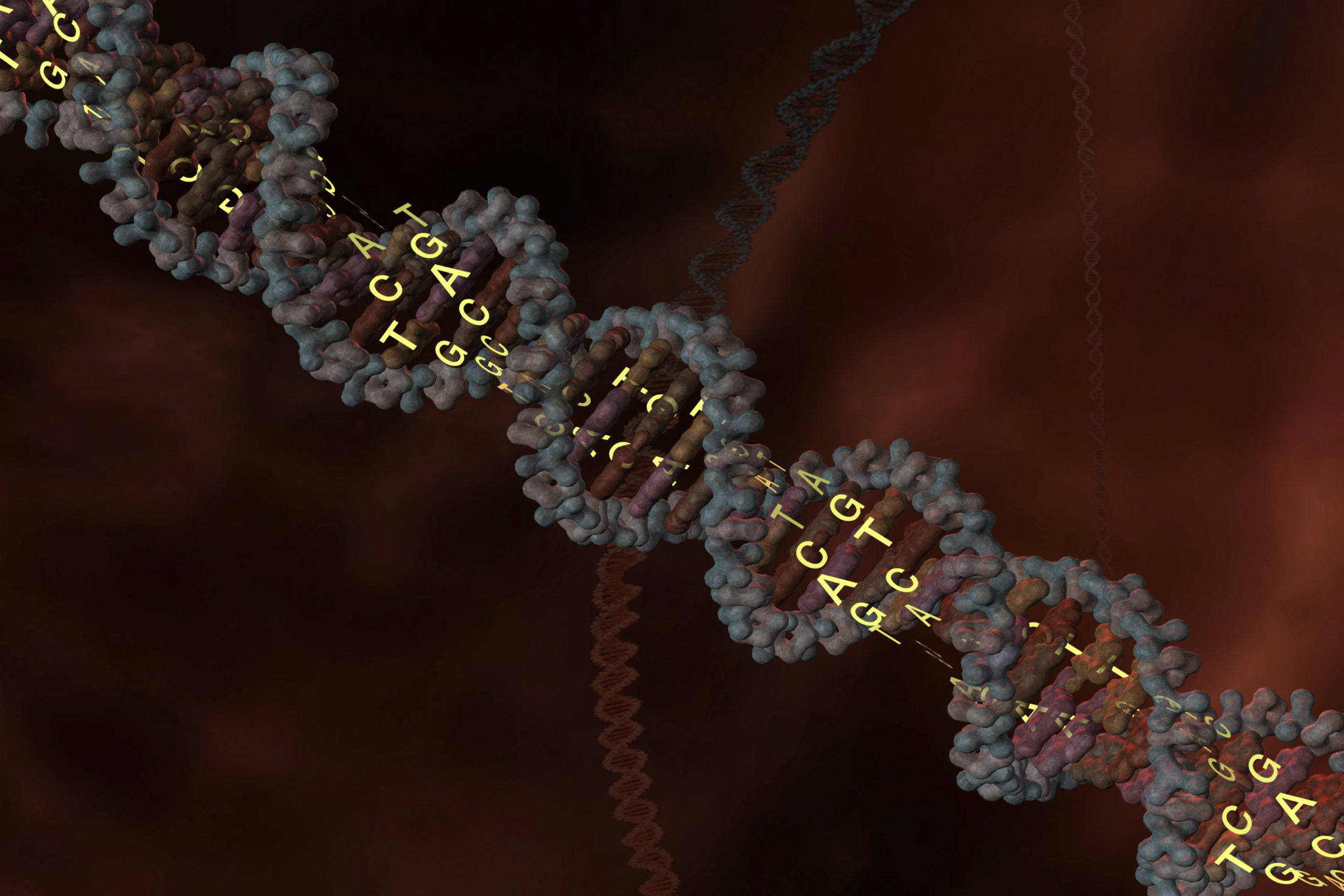
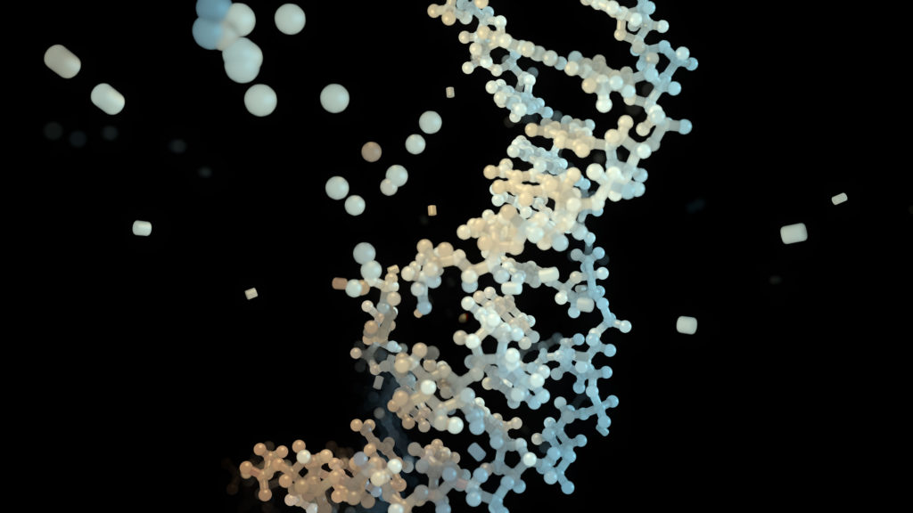
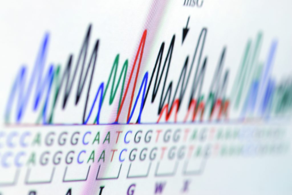
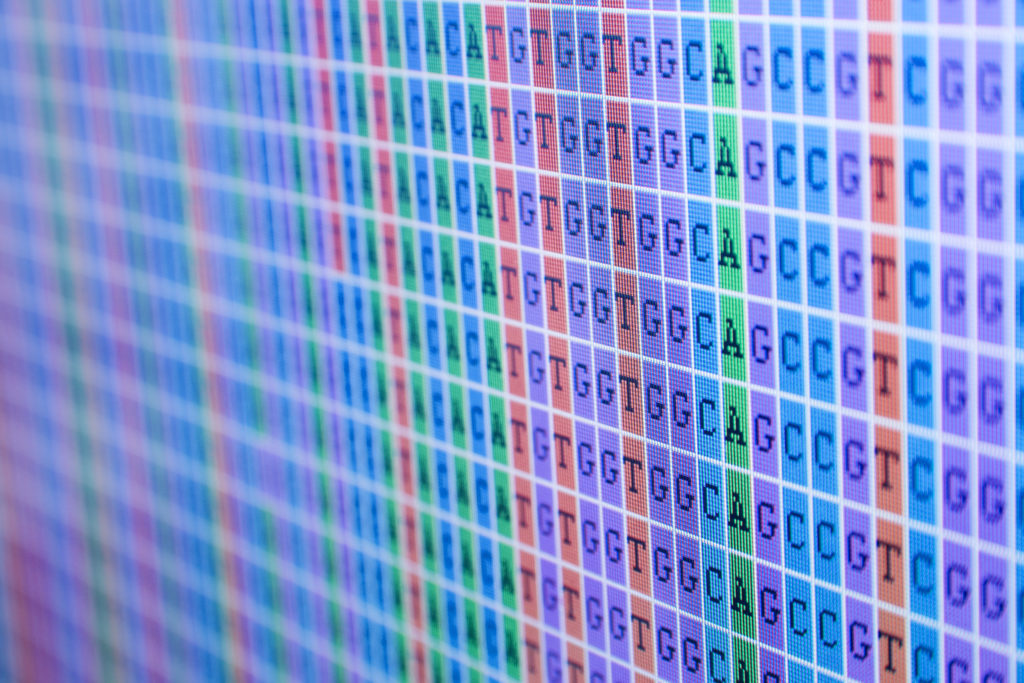
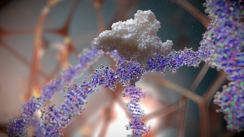
ACGT (adenine, cytosine, guanine, thymine)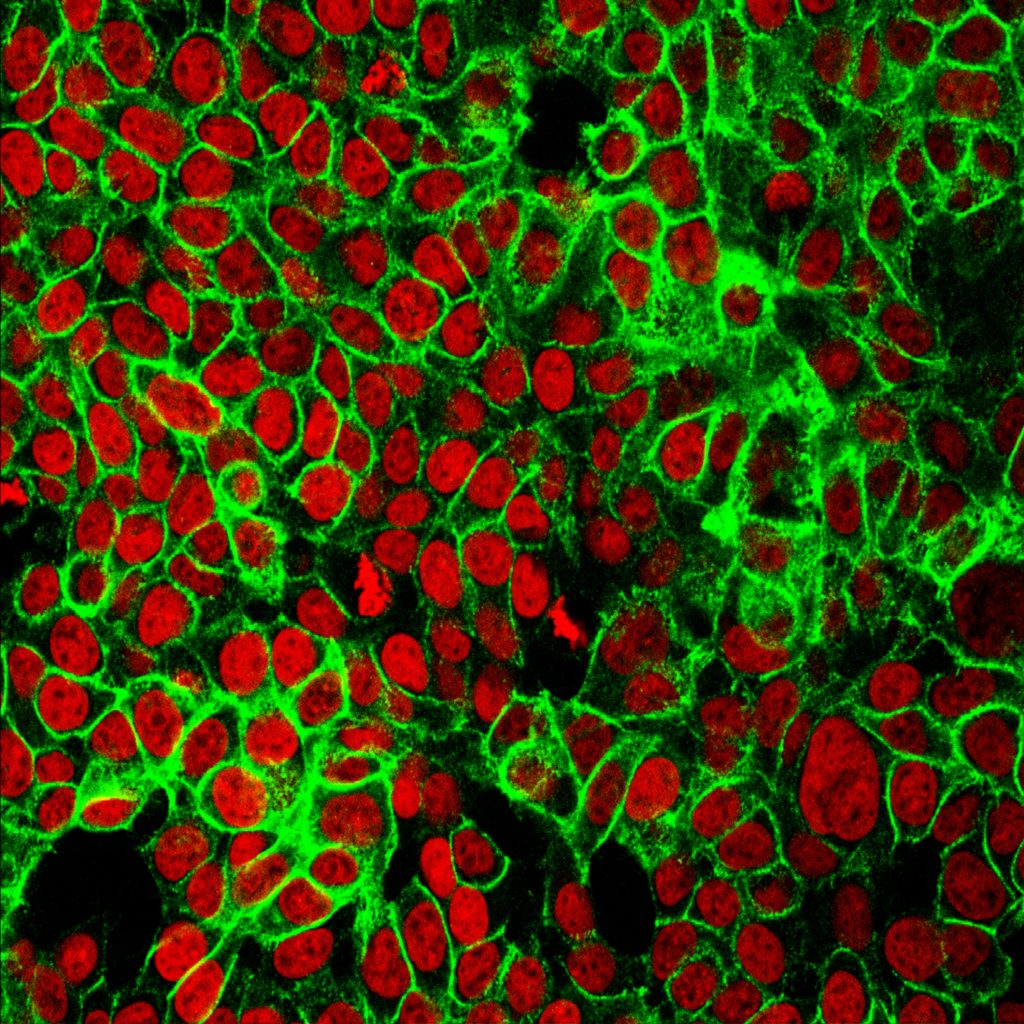 Photo by
 National Cancer Institute onUnsplash Photo by National Cancer Institute on Unsplash 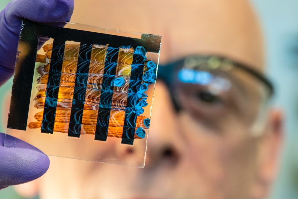 Photo by
 Science in HD onUnsplash NREL researchers have developed an interdigitated back contact solar cell design in which the metals and transport materials are solution processed by either ink jet or spray coating. Combined with a perovskite ink formulation with a low boiling point (<80 C) allows "paintable" solar cells.Photo by Science in HD on Unsplash  Photo by
 Michael Schiffer onUnsplash Photo by Michael Schiffer on Unsplash  Photo by
 National Cancer Institute onUnsplash Mapping the position of genes in the cell nucleus sheds light on basic principles governing the genome. Here, a single gene called Pem (purple) has been localized using fluorescence in situ hybridization. DNA is stained blue; the cell cytoplasm is stained green.  Photo by
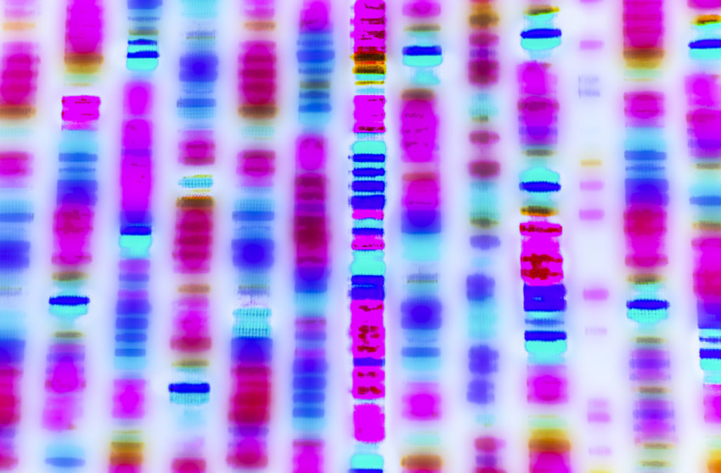
 National Cancer Institute onUnsplash A dye marker on agarose gel used to separate DNA.  Photo by
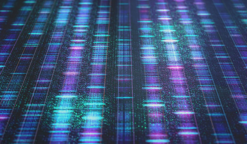
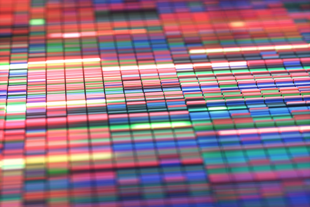
 National Cancer Institute onUnsplash Three-Dimensional Landscape of Genome.  Photo by
 National Cancer Institute onUnsplash A technician using a microtome, an instrument that cuts extremely thin sections of material for examination under a microscope. 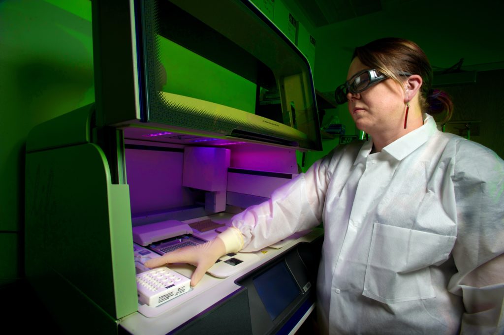 Photographer James Gathany
A whole genome DNA sequencer, in order to determine the “DNA fingerprint” of a specific bacterium. 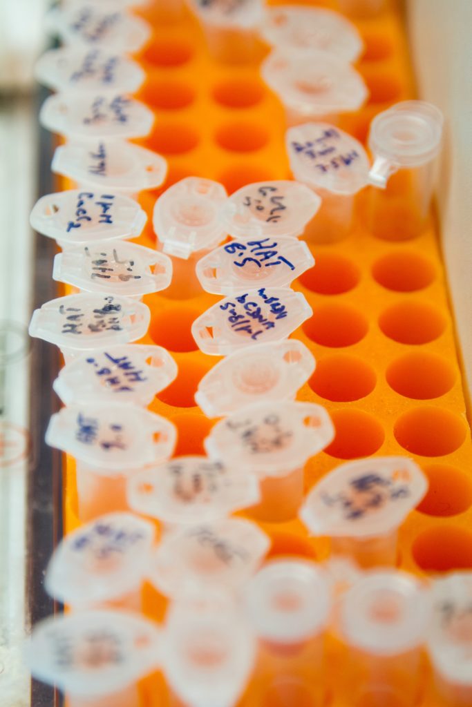 Photo by
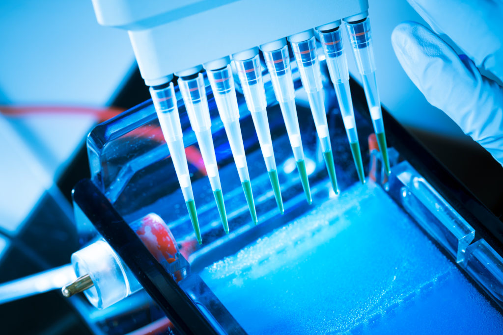
 National Cancer Institute onUnsplash Microcentrifuge tubes in a rack. 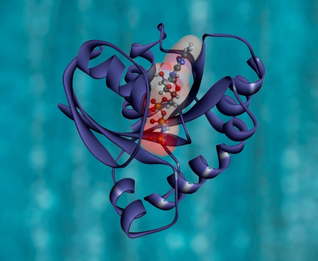 Photo by
 National Cancer Institute onUnsplash Photo by National Cancer Institute on Unsplash 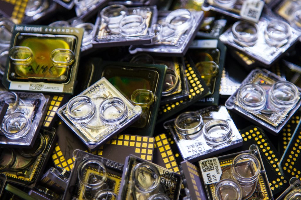 Photo by
 National Cancer Institute onUnsplash DNA Genotyping and SequencingA box containing used sequencing chips at the Cancer Genomics Research Laboratory, part of the National Cancer Institute’s Division of Cancer Epidemiology and Genetics (DCEG). 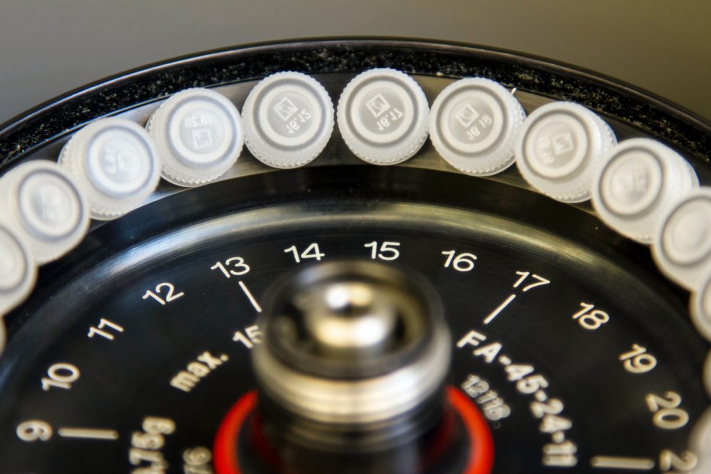 Photo by
 National Cancer Institute onUnsplash DNA Genotyping and Sequencing. Centrifugation of DNA samples prior to high-throughput genotyping and sequencing at the Cancer Genomics Research Laboratory, part of the National Cancer Institute’s Division of Cancer Epidemiology and Genetics (DCEG). 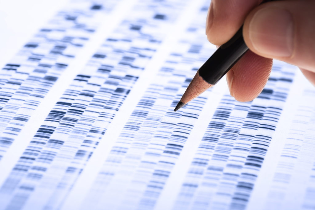 Wissenschaftler untersucht DNA-Gel
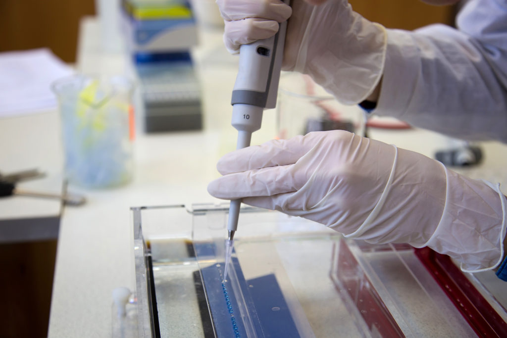
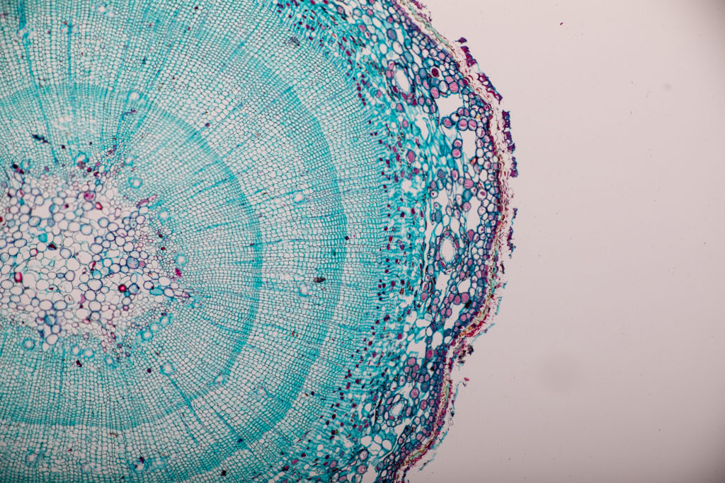
Cross-section Dicot, Monocot and Root of Plant Stem under the microscope. 
Cell structure 
Components of Eukaryotic cell, nucleus and organelles and plasma membrane. 
Bacteriophage attacking E. coli bacteria.